Detection of actin filaments in electron microscopy images
This project was executed by Tien Chen Lin from Frank Bradke’s Lab at DZNE Bonn in collaboration with Florian Schur’s Lab at IST Austria.
Objective and outcome
To detect actin filaments in an automated way, the raw images had to be cleaned from dirt, membrane fragments and other experimental artifacts.
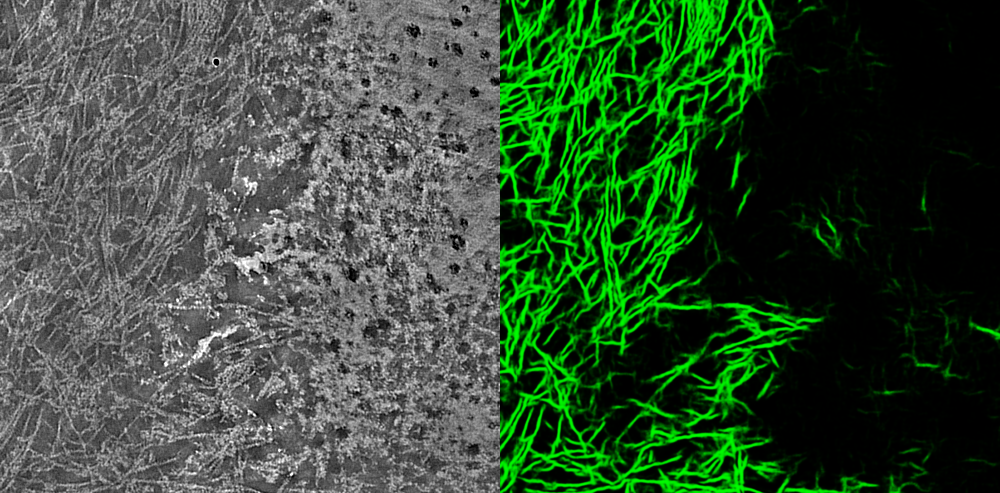
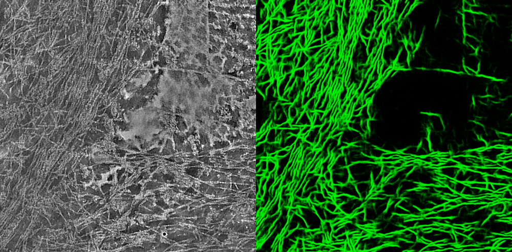
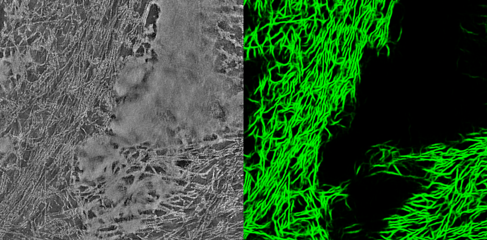
Tien Chen trained a model with YAPiC using unet_2d for detection of actin filaments (green). Membrane fragments and other experimental artifacts were defined as background.
Look at the three example images below: On the left, you see the raw input image. On the right you the the YAPiC output: Actin filaments (green) are enhanced and clearly separated from each other. Membrane fragments are filtered out.


How to proceed from here?
YAPiC is a tool for preprocessing your raw data, i.e, to make subsequent object detection both easier and more precise. However, object detection itself is not included in YAPiC. This could be done with thresholding and skeletonization in Fiji, or using more advanced tracing algorithms (e.g. on basis of localized radon transform)